Sunday, 27 December 2020
TYPES OF RIBOSOMES

Wednesday, 29 May 2019
FIT AND SLIMIZ: The need for exercise
FIT AND SLIMIZ: The need for exercise: The need for exercise (Why exercise?) Before we know why we should do exercise, it's important to know what exercise is. Many peopl...
Tuesday, 1 January 2019
RIBOSOMES

RIBOSOMES Ribosomes are small, dense,rounded and granular particles of the ribonucleoprotein.
DISCOVERY Ribosomes was studied by palade in 995 for the first time.
OCCURRENCE AND DISTRIBUTION: These are small granular structures found in prokaryotes and eukaroyets. They occur either freely in the matrix of mitochondria chloroplast and cytoplasm (i.e, cytoplasmic matrix) or remain attached with the membranes of the endoplasmic reticulum nucleus.
ROLE They carry put proteins synthesis by translating mRNA in the polypeptide chain.
CHEMICAL COMPOSITION
Ribosomes are composed of proteins and ribosomal RNA. Ribosomal RNA in eukaryotes is a four types and three types in prokaryotes. Ribosomes are formed in the nucleolus.

STRUCTURE:
These are small cytoplasmic granules ranging from 20nm-30nm in diameter. Ribosomes are consisting of two sub units of unequal size that fit closely together.
Ribosomes found in eukaryotic cell are 80S, and prokaryotic cell are 70S.

SUBUNITS The subunits are referred as the smaller subunits and the larger subunits. The larger subunits in eukaryotes is 60S while in prokaryotes is 50S. The smaller subunits in eukaryotes is 40S and in prokaryotes 30S.
SVEDBREG UNIT: "S" refers to Svedbreg unit that is the sedimentation rate in ultracentrifuguation.
EUKARYOTIC RIBOSOMES: Its smaller sub unit is 30S and Larger subunit is 50S and complex is 70S.
PROKARYOTIC RIBOSOMES: Its smaller sub unit is 40S and larger sub unit is 60S and complex is 80S.
ENDOPLASMIC RETICULUM

ENDOPLASMIC RETICULUM Endoplasmic reticulum is an extensive network of interconnected channels and vesicles called cisternae, these membranous channels extends from the nuclear membrane into the cytoplasm. The membrane enclosed space between the cisternae is called the lumen of ER.
TYPES OF ENDOPLASMIC RETICULUM On the basis of the presence or the absence of the ribosomes there are two types of ER
SMOOTH ENDOPLASMIC RETICULUM The surface of smooth ER is devoid of the ribosomes, and thus called the smooth ER.
FUNCTION OF SMOOTH ENDOPLASMIC RETICULUM:
AS A TRANSPORT SYSTEM The endoplasmic reticulum acts as an intracellular circulatory or transporting system. The channels help in the transport of material in the cell.
DETOXIFICATION OF TOXIC SUBSTANCE Smooth ER helps in the detoxification of toxic substance in liver
LIPID METABOLISM: Smooth ER helps in lipid metabolism.
ROUGH ENDOPLASMIC RETICULUM: The surface of the rough ER is studded with ribosomes and thus is called as rough endoplasmic reticulum. RER is in direct contact with the nuclear membrane, and RER the nuclear membrane is also studded with ribosomes.
FUNCTION OF ROUGH ENDOPLASMIC RETICULUM:
SKELETAL FRAMEWORK:
The endoplasmic reticulum provides an ultra- structural skeleton frame work to the cell and gives mechanical support to the colloidal cytoplasmic matrix.
AS A TRANSPORT SYSTEM:
The endoplasmic reticulum acts as an intracellular circulatory or transporting system.
The channels help in the transport of material in the cell.
PROTEIN SYNTHETIC MACHINERY:
Rough ER helps in the synthesis of proteins.
FORMATION OF NUCLEAR ENVELOP:
The ER membranes from the new nuclear envelope after each nuclear division.

Saturday, 29 December 2018
CHROMOSOMES

CHROMOSOMES
During cell division chromatin material becomes shorter and thicker forming chromosomes, chromosomes stains dark and under compound microscope appears to be made up of arms and centromeres.
Centromeres attached the two arms called chromatids. Centromeres has a proteins called kinetochore proteins through which chromosomes is attached to the spindle fibers during cell division.

COMPOSTION OF CHROMOSOMES:
A chromosomes is composed of DNA and proteins. (one chromosomes =one DNA molecules)

DIPLOID AND HAPLOID NUMBER OF CHROMOSOMES The full number of chromosomes in normal body cells is diploid (2n) whereas haploid (n) is the half number of chromosomes present in germ or gamete cells. For example, human sperms and eggs have 23 each and those of Drosophila have 4 each in sperms and eggs. So in this way after fertilization the number of chromosomes remains constant in the next generation
EUCHROMATIN: Euchromatin is the well dispersed form of chromatin which takes lighter DNA-stain and is genetically active, i.e. it is involved in gene duplication, gene transcription (DNA- dependent RNA synthesis) and morphological expression of a gene through some type of protein synthesis.
HETEROCHROMATIN: Heterochromatin is the highly condensed form of chromatin which takes dark DNA-stain and is genetically inert. Such type of chromatin exists both in the region of centromere (called constitutive heterochromatin) and in the sex chromatin (called facultative heterochromatin) and is late replicating one.
Friday, 28 December 2018
NUCLEUS

NUCLEUS
DISCOVERY
Nucleus was for the first time described by the English Robert Brown in 1831.
OCCUERENCE AND DISTRIBUTION
Nucleus is present in all eukaryotic cells. The nucleus is centrally located and roughly spherical cellular component which controls all the vital activities of the cytoplasm and carries the heredity material the DNA in it.
CELL WITH NO NUCLEUS:
Prokaryotic have no nucleus. Mammalian erythrocytes (red blood cells) lose their nuclei when they mature.
SIZE
It is typically 10 um in diameter.
IMPORTANCE
It is one of the most important organelle because it controls all the metabolic activities and his the genetic information in the form of chromosomes and DNA.

STRUCTURE OF NUCLEUS Nucleus consist of 1) Nuclear envelop 2) Nucleolus 3) Nucleoplasm and 4) Chromosomes NUCLEAR ENVELOP: The nucleus is bounded by two membranes, which together called nuclear envelope. The outer membrane of the nuclear envelope is continuous with the endoplasmic reticulum. NUCLEAR PORES: Over the surface of the nuclear envelope, are shallow depressions called nuclear pores. These pores helps in the transport of the proteins into the nucleus and mRNA from nucleus into the cytoplasm. NUMBER OF NUCLEAR PORES: Egg cells have maximum number of pores, while minimum in RBCs. NUCLEOLUS: It is a granular body with in the nucleus and is not bounded by a membrane. There may be one or more nucleoli in the nucleus. REGIONS OF NUCLEOLUS: It is composed of two regions; the peripheral granular area composed of precursor which helps in synthesis of ribosomal sub units, and the central febrile area consisting large RNA and DNA. NUCLEOPLASM: The space between the nuclear envelope and the nucleolus is filled by a transparent, semi- solid granular ground substance or the matrix known as the nucleoplasm It is a colloidal mixture of organic and inorganic salts and ions. FUNCTION OF NUCLEOPLASM: Nucleoplasm houses nuclear content. It also serves as storage place for enzymes, raw material needed for DNA replication and synthesis of RNA. ROLE OF NUCLEUS: RNA SYNTHESIS: RNA is synthesized and stored in the nucleolus. FORMATION OF RIBOSOMES: Proteins imported from the cytoplasm are assembled with RNA forming ribosomal sub units. These sib units then exit the nucleus through the nuclear pores to the cytoplasm, where a large and a small sub units can assembled into a ribosome.
THE ROLE OF PROTEINS, LIPIDS AND CHLORESTOL
THE ROLE OF PROTEINS, LIPIDS AND CHOLESTEROL

PROTEINS AND THEIR ROLE: TRANSPORT: Transmembrane proteins from water channels through which the transport of material and ions takes place. CELL RECOGNITION: Extrinsic proteins help in the recognition of the cell and foreign particles.
ADHESION:
Some of the proteins help in the adhesion of cell to each other.

ROLE OF LIPIDS:
AS ANTI-FREEZE:
Unsaturated Fatty acid tails helps in protecting the cell membrane from freezing.

ROLE OF CHOLESTEROL *) Cholesterol prevents the plasma membrane from overexpansion. ROLE OF GLYCOLIPIDS AND GLYCOPROTEINS: *) It helps in the formation of glycocalyx, which in some bacteria facilitate attachment to the substrate. Furthermore acts as hormone receptors. FUNCTION OF PLASMA MEMBRANE: CELL SHAPE: Plasma membrane gives specific shape to the cell. PHAGOCYTOSIS: Plasma membrane helps in phagocytosis and pinocytosis EXCREATION: Plasma membrane helps in the excretion of the waste materials from the cell, (urea,uric acid etc.) CELLULAR SECRETION: Plasma membrane helps in the secretion of useful substances (hormones, enzymes, etc.) Plasma membrane prevents the escape of cytoplasmic organelle from the cell.
PLASMA MEMBRANE

PLASMA MEMBRANE
OCCURRENCE OF PLASMA MEMBRANE
Plasma membrane or the cell membrane is usually found in all types. it is the outer most boundaries of the animals cells and inner to cell wall in plant cells.
CHEMICAL COMPOSITION OF PLASMA MEMBRANE
Cell membrane is chemically composed of lipoproteins as lipids (20-40%) and proteins (60-80%). In addition there is small quantity of carbohydrates and cholesterol is also present.
lipids present are phospholipids and sterol, the cholesterol in the animals cell are sterol and in that of plant cell are phytosterol.

STRUCTURE AND MORPHOLOGY: The most acceptable model for the membrane structure was proposed by singer and Nicholson in 1972, this model is called as the fluid Mosaic model. Fluid part refers to the phospholipids molecules and mosaic part refers to the proteins. THE LIPIDS BILAYER (FLUID) The membrane is composed of a lipid bilayer in which the proteins are sandwiched in a mosaic pattern. The outer surface of the lipids layer is hydrophilic and is glycerol. The inner surface is hydrophobic and is composed of water- insoluble fatty acid tail. ARRANGEMENT OF PROTEINS (MOSAIC) The protein does not form a layer on lipids bilayer layer but are arranged in a mosaic pattern. TYPES OF PROTEINS: There are two types of proteins. EXTRINSIC PROTEINS: The proteins that lie on the outer surface of the lipid bilayer are called the extrinsic proteins or perpheral proteins most of which are present on the cytoplasmic side. INTRINSIC PROTEINS: The proteins that are completely buried in the lipid layer are called the intrinsic proteins.
THE GLYCOCALYX
Carbohydrates attached to the proteins forms glycoproteins, and carbohydrates attached to the lipids forms glycolipids. These carbohydrates form a layer on above the membrane called glycocalyx

Thursday, 27 December 2018
CELL WALL

CELL WALL
DISCOVERY The cell wall was discovered by Robert Hooke in 1665.
OCCURRENCE It is found in all plant cells, fungi, algae and bacteria, but absent in animals cell and protozoa. it is the outer most boundaries of the plant cells.
CHEMICAL COMPOSITION Cell wall is composed of micro fibrils, these fibrils are cemented together by lignin pectin and other substances. Chemical composition of cell wall varies depending on the type of cell.
CELL WALL IN BACTERIA: Bacterial cell wall is chemically composed of peptidoglycan.
CELL WALL IN PLANTS:
Cell wall of plants cell is composed of cellulose, hemicellulose, pectin and lignin.
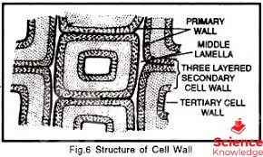
STRUCTURE OF CELL WALL The cell waLls have three fundamental parts.
MIDDLE LAMELLA Middle lamella joins together the two adjacent cells. it is usually thin and about 1 um in thickness. Middle lamella is formed during cytokinesis by the cell plate during cytokinesis,
CHEMICAL COMPOSITION: In woody tissue it is mainly composed of magnesium and calcium pectate. Middle lamella is commonly lignified.
PRIMARY WALL Primary wall is the first wall formed during development. it is usually 1-3 um in thickness.
CHEMICAL COMPOSITION: It is composed of cellulose, pectin mostly polysaccharides and hemicellulose.
SECONDARY WALL It is laid down inside the primary wall. It is usually thick about 5-10 um. It is more or less rigid, both the wood and bark are the secondary cell walls. Secondary cell wall give rigidity to the plant.
CHEMICAL COMPOSITION: Secondary cell wall is composed of hemicellulose (xylan) and lignin minerals (Ca,Mg)
PLASMODESMATA:
Plasmodesmata are small pores in the cell wall. Cells interact though plasmodemata with the protoplasts of adjacent cells across the wall.

IMPORTANCE OF CELL WALL/FUNCTIONS OF CELL WALL *) Cell wall gives strength and rigidity to the cell. *) Cell wall prevents the cell from osmotic lysis. *) Cell wall limits the entry of larger molecules inside the cell. *) Cell wall is porous and allow free passage of water and dissolved minerals.
SPECTROPHOTOMETRY, MICRO DISSECTION AND MICROMETRY

SPECTROPHOTOMETRY
DEFINITION
Spectrophotometry is the measure of amount of light that passes through the sample can be calculated that how much light was absorbed by the sample.
SPECTROPHOTOMETER
A spectropohtometer is a photometer that can measure intensity of wave length of light source.
OPTICAL DENSITY
Ability of specimen to absorb or block the passage of light.
FACTORS
The cell mass is directly proportional to the optical density. The amount of light passes through the solution is indicative of the concentration of certain chemicals that do not allow light to pass through.
APPLICATIONS
Spectrophotometry can be used to determined the wave length of light that take part 1 photosynthesis.

MICRO DISSECTION
It refers to a variety of techniques in which microscope is used for the dissection of cells or its organelles.
TYPES
Different kinds of techniques involve in micro dissection.
*) CHROMOSOME MICRO DISSECTION
To remove a portion from a complete chromosomes, a fine glass needle is being used under a microscope.
*) LASER MICRO DISSECTION
Use of a laser through a microscope for the dissection of selected cells.
*) LASER CAPTURE MICRO DISSECTION
Use of a laser through a microscope to cause selected cells to adhere to a film is called laser capture micro dissection.

MICROMETRY
DEFINITION Measuring the size of microscopic objects is called micrometry. This can be done by using specially designed scales. (1) OCULAR MICROMETER One of the scales is placed in eyepiece called ocular micrometer. (2) STAGE MICROMETER Other on the stage (stage micrometer) The micrometer have equally spaced divisions. Before using the eyepiece micrometer to measure a particular structure, you will have to find out the real width of each unit on the scale at each magnification. In other words you have to calibrate the micrometer. This can be done by replacing the specimen with the stage micrometer, and to measure the eyepiece units at each magnification.
GEL ELECTROPHORESIS

GEL ELECTROPHORESIS
DEFINITION:
The techniques which is used for the separation of molecules on the bases of their size and charge in gel under the influence of electric field is called gel electrophoresis.

PROCEDURE: In this techniques agarose gel act as molecular sieve and separate the molecules on the basis of their size and surface charge. The gel is poured into the plastic plates to form a viscous slab. Two ends of the slab are suspended in salt solution which are connected b electrodes to a power source. When voltage is applied the molecules start moving through the gel. Negatively charged molecule moves towards positive pole and positively charged molecules moves towards the negative pole.
TYPES OF ELECTROPHORESIS
*) ANAPHORESIS: (Separation of histone)
*) CATAPHORESIS: (Separation of DNA/RNA)
FACTORS AFFECTING THE SPEED OF MOLECULES: Two factors affect the speed, with which charged molecule move towards an electrode. a) The amount of charge (greater charge, faster movement and vice versa) b) The size of molecule (smaller molecules, faster movement and vice versa) c) Shape of DNA d) Concentration of gel.
TYPES OF GEL:
Different gel types and conditions are used for different molecules and types of applications.
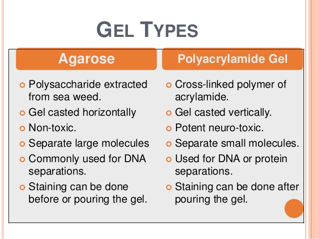
AGAROSE: A common gel for separating large DNA fragments is made of agarose
POLYACRYLAMIDE: For separation of small DNA fragments polyacrylamide gel is used.
APPLICATION: Gel electrophoresis separates macro molecules such as Nucleic Acid and proteins on the basis of their rate of movement through an agarose gel in an electric field. The distance a DNA molecule travels is inversely proportional to its length or size.
Wednesday, 26 December 2018
CHROMATOGRAPHY

CHROMATOGRAPHY Chromatography is the separation of sample components of a mixture based on differential affinity for a mobile versus a stationary phrase. separation involves two phrase.
MOBILE PHASE The mobile phase is a liquid or gas with a mixture of substances to be separated.
STATIONARY PHASE The stationary phase is a solid substance through which the mobile phase moves. The separation depends on chemical and physical properties of the molecules such as solubility and molecular mass. ION EXCHANGE AND ABSORPTION CHROMATOGRAPHY When the components are separated by ionic bonds the chromatography is ion exchange, and when the components are absorbed by stationary phase it is known as absorption chromatography.
PAPER CHROMATOGRAPHY It is used for the separation of photosynthetic pigments, sugar or amino acids. In this type of chromatography paper is used as a stationary phase.
PROCESS *) A mixture is dissolved in a liquid and spotted near one end of a paper strip. *) The end of the paper nearest to spot is placed in the solvent in such a way that alcohol does not touch the spot. *) Alcohol which moves through the paper by capillary action carrying the molecules with it, and results in the formation of bands on the paper.
DIAGRAM

TYPES
COLUMN CHROMATOGRAPHY
In column chromatography the stationary phase is a supporting phase is a supporting tube, with a paper or silica on the inner surface of the tube.

THIN LAYER CHROMATOGRAPHY (TLC)
When the stationary phase is cellulose or thin silica then it is called as thin layer chromatography. .

TISSUE CULTURE

TISSUE CULTURE
DEFINITION The in-vitro culturing of cell, tissue or organ of an organisms in highly aseptic environment on artificial medium is called tissue culture. Plants cells unlike most of the animals cells have copies of many functional genes and thus they can be propagated from a cell, and have the same genotype (genetically identical plants.)
HOW TISSUE CULTURE IS DONE

APPARATUS

*) Centrifuge
*) Water bath
*) Sterilizer
*) Liquid nitrogen freezer
*) Hemacytometer, this device is used for counting cells
*) Incubator, a device that maintains humididty, temperature and gas concentration.
*) Cell culture hood that provides a hygienic environment.
*) Broth, or agar growth medium

PROCEDURE
a) PROCUREMENT OF EXPLANT: Plant tissue cultures are initiated from explants, which are complete organs or small sections of tissue or a single cell removed from an intact plant.
b) STERILIZATION: Before tissue culture, glassware and explants are sterilized in chemical solutions (sodium or calcium hypochlorite, solutions). c) PREPARATION OF A GROWTH MEDIUM: A suitable growth medium is prepared with all the essential element, organic nutrients, vitamins and plant hormones.
d) INOCULATION: Explant are allowed to grow in growth medium. Inoculation is performed in laminar flow hood that provides a germ free-sterile environment.
e) DEVELOPMENT OF CALLUS: The growing explants from a mass of undifferentiated cells called callus.
f) DEVELOPMENT OF PLANTLETS: As roots and shoots appear from tghe callus these are planted in soil to produces individual plants, due to chnages in hormones concentration.
Tuesday, 25 December 2018
WHAT IS CELL

CELL
DEFINITION
The cell is the basic structural and functional unit of all living organisms. it is the smallest unit that can carry out all activities of life.

TYPES OF CELL There are two main types of cells; Prokaryotic cell and Eukaryotic cell. Eukaryotic cells have a proper nucleus and double membrane- bounded organelle. Prokaryotes in contrast, do not have a nucleus. They have a region called nucleoid in which the genetic material is located
CELL HISTORY Ancient Greek philosophers such as Aristotle concluded that "all animals and plant are constituted of a few elements which are repeated in each of them." The elements now days called cells Further growth and development of cell biology is associated with the development of optical lenses and to the combination of these lenses in the construction of the compound microscopes. Robert Hooke (1635-1703) was proposed the term cell (Latin, Cella = holloow space) in 1665 He examined a thin slice cut from a peice of dried cork under the compound microscope which were built by him by using 30 multiply microscope. Leeuwenhoek was the first to observe free-living cells such as human sperm cells etc. Robert Brown (1773-1858) discovered and named the nucleus in plant cells.n In 1838, a German botanist Mathias Jacob Schleiden (1804-1881) put forth the idea that cells were the units of structure in the plants. In 1839, his coworker a German Zoologist Theodor Schwann (1810-1882) applied Schleiden's idea to the animals.Later, both of them postulated that the cell is the basic structural and functional unit of all living organisms.
DIAGRAM


CELL THEORY
Credit for developing cell theory is usually given to three scientists: Theodor Schwann, Matthias Jacob Schleiden, and Rudolf.
MAIN FEATURES
This theory has the following points.
*) All living organisms are composed of one or many cells.
*) All new cells arise from division of pre-existing cells.
*) Cells contain the hereditary material of an organisms which is passed from parent to daughter cells.
*) All metabolic processes take place within cells.
*) Basic chemical compostion of cells within similar species is same.

HOW ARE THE CELLS STUDIED There are several different techniques to study cells such as: Microscopy (includes simple light microscope, compound light microscope) differential staining, centifugation, cell/tissue culture,chromatography, electrophoresis etc. THE EARLIEST MICROSCOPE The simple microscope was invented by Antony Van Leeuwenhoek in 1670. This microscope was composed of only one lense and able to magnify an object up to 250 times. Robert hook developed a microscope called the compound microscope with two set of lenses called the ocular lens and the objective lens, in this type the image magnified by the objective lense is magnified a second time by the ocular lens. These microscope rely on the light.
Sunday, 23 December 2018
WHAT IS BIOLOGY

BIOLOGY
DEFINITION
The word biology is derived from two Greek words, bio means life, and logos means to study or thought "Biology is the scientific study of life or living organisms"

WHAT IS ORGANISM All living thing are organisms 1.1 FUNDAMENTAL CHARACTERISTICS OF LIVING ORGANISMS
ORGANIZATION
All organisms are composed of one or more cells with DNA present in the nucleus in case of Eukaryotes and within a cytoplasm in prokaryotes

REPRODUCTION
The process of production of young ones of their own types for the survival of their race is called reproduction. All organisms can produce their young ones,

GROWTH AND DEVELOPMENT Growth and development is the main characteristic of living organisms all organisms either plant or animals reproduce, grow and develop ADAPTATION Usually, all organism has the ability to adjust to the environment.They have the ability to adapt to the certain environment and thus evolve with the passage of time
METABOLISM The sum of the all the biochemical activity is called metabolism. For sustainability and viability, organisms need to be metabolically active
Subscribe to:
Posts (Atom)